Information
These codes make up the framework for segmenting the doppler ultrasound scans.
Table of Contents
Introduction to Ultrasound Segmentation Package (usseg)
This documentation introduces the Ultrasound Segmentation Package (usseg), a specialized toolkit for extracting and processing data from Doppler ultrasound images. Developed with a focus on compatibility with the Voluson E8 ultrasound system.

The package is composed of a series of interconnected functions, each designed as a step in the sequential process of ultrasound image segmentation and analysis. While it offers particular advantages for users of the Voluson E8, there is scope to adapt these tools for use with other ultrasound systems.
The purpose of this documentation is to provide an overview of the package, offering insights into:
The mechanism and workflow for processing images.
The functionalities and applications of each tool within the package.
Example usage for a series of situations, such as single image processing, batch processing, and developer related cases.
For detailed information about individual functions, please refer to the respective sections linked here. This package, being open-source, is part of a collaborative effort to advance ultrasound imaging technology, and we welcome contributions and suggestions from the community.
Overview
The Ultrasound Segmentation Package facilitates two primary blocks of functionality: text extraction from images and ultrasound image segmentation. Each block consists of a sequence of functions designed to work independently yet contribute collectively to the overall process.
Text Extraction Process
colour_extract_vectorized: Filters the image to highlight a specific target colour of pixel, preparing it for text extraction.

Text_from_greyscale: Processes the filtered image to extract text, matching lines to specific target words.
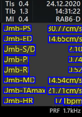
Metric_check: Performs a common-sense check on the extracted measurements using knowledge of their interdependencies and known physiological limits, ensuring data accuracy.

Following the successful extraction and validation of text data, the workflow transitions to the image segmentation process.
Image Segmentation Process
Initial_segmentation: Begins with a coarse segmentation of the waveform.
Define_end_ROIs: Defines regions adjacent to the coarse segmentation.

Segment_refinement: Refines the segmentation within the coarse boundaries.

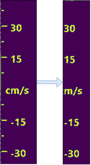
Search_for_ticks: Identifies ticks in the axes ROIs for accurate scaling.
Search_for_labels: Locates labels within the axes ROIs for data extraction.
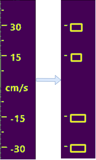
Plot_Digitized_data: Digitizes the extracted data to plot the waveform.
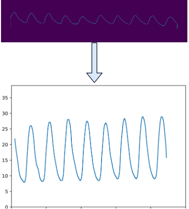
Plot_correction: (Optional) Adjusts the time axis based on heart rate data.
Annotate: Visualizes the segmentation steps on the original image.

Each function in these sequences plays a vital role in the overall process, which aims for accurate data extraction. For more in-depth information about each function, please refer to the detailed descriptions in the here section of this documentation.
Usage Examples
Some common usage examples include segmenting a single image, or processing a batch of images.
Processing a single image
For processing a single image, the data_from_image function is imported and provided with PIL and cv2 versions of the image. This could be done through the following code:
# Module imports
import numpy as np
from PIL import Image

# Local imports
from usseg import data_from_image

img_path = "Path/to/a/ultrasound/image.JPG"

PIL_image = Image.open(img_path)
cv2_image = np.array(PIL_image)
df, (xdata, ydata) = data_from_image(PIL_image, cv2_image)
Alternatively, the single image processing script within the tests folder can be modified for any given input image.
Batch processing images
For processing groups of images, the usseg.main module can be used. This module is designed to process a series of images contained in a folder and save to specified location, defined in the config.toml. A visualisation file output.html is generated for evaluation the data extraction. This can be done through:
python usseg/main.py
Limitations and future work
Work on this data extraction package is ongoing. Testing and development have highlighted several limitations and areas for improvement, guiding the next steps in its evolution.
The package is currently designed for scans typical of the Voluson E8. This typically includes measurements in the upper right corner in yellow and the Doppler waveform in the center bottom half, including both left and right y-axes. While the functions leverage these features to ensure accuracy, a limitation is their ineffectiveness with scans from different types of machines. However, the framework is robust, and minor modifications could adapt it for other ultrasound devices.
Regarding data extraction, there are occasions where text data is not correctly extracted. Errors might include missing decimal points or misinterpretation of characters (e.g., ‘5’ read as an ‘S’). Common-sense checks help mitigate these errors, but they are not foolproof.
Waveform segmentation and digitization quality often depend on the image’s contrast. Faint waveforms against the black background might result in inaccurate segmentation or complete failure to identify. Furthermore, some scans are inverted, and although there are measures to adjust these during digitization, exceptions can still occur.